
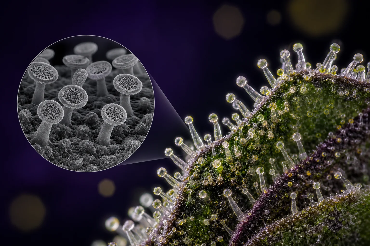
Macro to micro: female inflorescences bear the stalked glandular trichomes that concentrate cannabinoids and terpenes; SEM inset shows the head’s discoid secretory cells.
Mainezilla original editorial visual · AI-assisted art directionWhat growers and scientists mean by 'trichome'
In everyday horticulture a trichome is simply a hair or hair‑like outgrowth of the epidermis. Botanically, however, 'trichome' is a morphological category that includes both non‑secretory hairs and complex glandular structures specialized for metabolite production. On Cannabis, both types exist and perform different functions: non‑glandular trichomes contribute to physical protection, while glandular trichomes synthesize and sequester secondary metabolites such as cannabinoids and terpenes. ([pmc.ncbi.nlm.nih.gov](https://pmc.ncbi.nlm.nih.gov/articles/PMC4740396/))
Because growers tend to care about measurable molecules, the shorthand 'trichome' commonly refers to glandular trichomes — the microscopic factories that accumulate THCA, CBDA, and a host of terpenes. But that shorthand hides important diversity: glandular trichomes differ in size, cell composition, and biochemical capacity. Recognizing that distinction helps when you read a microscopy study, a proteomics paper, or a lab certificate that reports 'flower' concentrations without specifying tissue. ([pmc.ncbi.nlm.nih.gov](https://pmc.ncbi.nlm.nih.gov/articles/PMC8488169/))
In this article I use 'glandular trichome' to mean the secretory, metabolite‑producing structures on female inflorescences and 'non‑glandular' to mean the protective or tactile hairs that are not primary sites of cannabinoid biosynthesis. Where the literature is ambiguous I point to the original microscopy or biochemical study. ([pmc.ncbi.nlm.nih.gov](https://pmc.ncbi.nlm.nih.gov/articles/PMC10485653/))
The three classical types: bulbous, sessile, and stalked
Since early scanning electron microscopy work, researchers have categorized Cannabis glandular trichomes into three broad morphologies: bulbous (very small), sessile (capitate‑sessile, short stalk), and capitate‑stalked (long stalk with large head). These categories remain useful because they correlate with head size, number of secretory disc cells, and—importantly—the capacity to accumulate cannabinoids. ([pmc.ncbi.nlm.nih.gov](https://pmc.ncbi.nlm.nih.gov/articles/PMC10485653/))
Morphologically, stalked glands often have a multicellular stalk, a short constricted neck, and a multicellular head composed of a stipe and a discoid arrangement of secretory disc cells; sessile glands are similar but smaller and with fewer disc cells; bulbous glands are the smallest and have only a few cells composing the head. These structural differences map to measurable differences in metabolic output in several studies. ([pmc.ncbi.nlm.nih.gov](https://pmc.ncbi.nlm.nih.gov/articles/PMC8016307/))
Importantly, the three‑type scheme is descriptive, not prescriptive: in developing bracts you can find a continuum of sizes and intermediate forms, and some sessile glands appear to be developmental precursors of stalked glands. Count and type will vary with genotype and inflorescence age. ([pmc.ncbi.nlm.nih.gov](https://pmc.ncbi.nlm.nih.gov/articles/PMC10071647/))
- Bulbous: smallest, few cells, low cannabinoid content per unit. ([pmc.ncbi.nlm.nih.gov](https://pmc.ncbi.nlm.nih.gov/articles/PMC8488169/))
- Sessile (capitate‑sessile): moderate size; historically considered intermediate. ([pmc.ncbi.nlm.nih.gov](https://pmc.ncbi.nlm.nih.gov/articles/PMC10485653/))
- Stalked (capitate‑stalked): largest heads, more disc cells, generally highest cannabinoid accumulation. ([pmc.ncbi.nlm.nih.gov](https://pmc.ncbi.nlm.nih.gov/articles/PMC10071647/))
Anatomy of the head and the stalk: cell types and compartments
Zooming into a stalked capitate trichome reveals a hierarchy of tissues. The multicellular stalk anchors the trichome to the epidermis; immediately above are stipe cells that subtend a discoid array of secretory disc cells (the 'head'). The secretory disc cells are the localized biosynthetic engines; many studies also report a large extracellular (apoplastic) storage cavity beneath the cuticle where synthesized metabolites accumulate. ([pmc.ncbi.nlm.nih.gov](https://pmc.ncbi.nlm.nih.gov/articles/PMC8016307/))
Disc cells differ from surrounding epidermal cells in subcellular organization: they have dense cytoplasm, abundant endoplasmic reticulum and plastids tailored to the pathway, and in some studies lack active chloroplasts that appear in supporting stipe cells. That separation — metabolic machinery in disc cells, photosynthetic elements in supporting cells — is functionally meaningful for precursor supply and localized biosynthesis. ([pmc.ncbi.nlm.nih.gov](https://pmc.ncbi.nlm.nih.gov/articles/PMC8016307/))
Biochemically, the head is a micro‑compartment where fatty‑acid‑derived and polyketide pathways converge; the final products (cannabinoid acids) are often transported or secreted into the storage cavity. Proteomics and transcriptomics of isolated trichome heads have begun to map the enzymes and transporters enriched in these cells. These molecular maps are why isolated‑head studies are more informative about biosynthesis than bulk‑flower analysis. ([pmc.ncbi.nlm.nih.gov](https://pmc.ncbi.nlm.nih.gov/articles/PMC8016307/))
Female inflorescences: where most glandular trichomes live
Glandular trichomes concentrate on female floral tissues — bracts and calyxes — where they are associated with reproductive structures. Female inflorescences show higher densities of glandular trichomes than leaves or male flowers, reflecting tissue‑specific developmental programs and the plant’s ecological investment in protecting and signaling around reproduction. ([pmc.ncbi.nlm.nih.gov](https://pmc.ncbi.nlm.nih.gov/articles/PMC10485653/))
Spatial distribution matters: bract tissues are densely covered, and within a single flower trichome density and maturation can vary. Trichome counts taken from different parts of the flower or from different developmental stages will therefore report different concentrations even for the same plant—this is a common source of apparent variation between lab certificates or research reports. ([pmc.ncbi.nlm.nih.gov](https://pmc.ncbi.nlm.nih.gov/articles/PMC10071647/))
Genotype and ontogeny both shape the trichome landscape. Some cultivars (genotypes) naturally make more stalked glands; others maintain a greater proportion of sessile or bulbous glands. During flower maturation the relative abundance often shifts toward larger, stalked glands. That dynamic explains why harvest timing and sampling protocol are crucial in comparative studies. ([pmc.ncbi.nlm.nih.gov](https://pmc.ncbi.nlm.nih.gov/articles/PMC10071647/))
Microscopy, isolation, and the limits of visualization
Understanding trichome anatomy depends on the microscope. SEM (scanning electron microscopy) provides surface morphology and is how the original three‑type classification was formed; light microscopy and confocal imaging reveal subcellular autofluorescence and organelle distribution; and transmission EM can show internal membranes and vesicles. Each technique has tradeoffs in resolution, sample preparation, and the risk of artifact. ([pmc.ncbi.nlm.nih.gov](https://pmc.ncbi.nlm.nih.gov/articles/PMC10485653/))
Because heads are small and chemically rich, isolation techniques (mechanical separation, centrifugation, or laser capture microdissection) paired with proteomics or transcriptomics allow molecular inventories of disc cells versus stalk or epidermal tissues. Proteomics of purified heads has identified enzymes and supporting proteins enriched in secretory tissues, giving a functional complement to imaging. ([pmc.ncbi.nlm.nih.gov](https://pmc.ncbi.nlm.nih.gov/articles/PMC8016307/))
But beware: any visualization or isolation method samples a subset of reality. SEM images represent fixed, dehydrated surfaces; biochemical isolation can bias toward more robust or adhesive heads. That’s why multi‑modal approaches—combining imaging, molecular profiling, and careful developmental staging—give the most reliable picture. ([pmc.ncbi.nlm.nih.gov](https://pmc.ncbi.nlm.nih.gov/articles/PMC8016307/))
- SEM: excellent for surface morphology; can distort soft structures. ([pmc.ncbi.nlm.nih.gov](https://pmc.ncbi.nlm.nih.gov/articles/PMC10485653/))
- Confocal/fluorescence: shows living autofluorescence and organelles when sample prep preserves them. ([pmc.ncbi.nlm.nih.gov](https://pmc.ncbi.nlm.nih.gov/articles/PMC8016307/))
- Proteomics/transcriptomics of isolated heads: reveals enzymes and transporters enriched in secretory tissue. ([pmc.ncbi.nlm.nih.gov](https://pmc.ncbi.nlm.nih.gov/articles/PMC8016307/))
From cell to molecule: where cannabinoids and terpenes are made and stored
Pathway localization studies converge on the secretory disc cells and the subcuticular apoplastic cavity as the principal sites of cannabinoid biosynthesis and accumulation. Core enzymes for the cannabinoid pathway are enriched in glandular tissues, and metabolites are detected at much higher concentration in isolated heads than in surrounding tissue. That concentration gradient underlies why head‑level studies are essential for mechanistic claims about production. ([pmc.ncbi.nlm.nih.gov](https://pmc.ncbi.nlm.nih.gov/articles/PMC8488169/))
Recent work has also highlighted the role of connected metabolic pathways — for example, oxylipin and fatty acid metabolism — in supplying precursors and modulating flux through the cannabinoid pathway. These findings underscore that trichome metabolism is integrated with broader cellular networks, not an isolated 'black box'. ([pubmed.ncbi.nlm.nih.gov](https://pubmed.ncbi.nlm.nih.gov/40343123/))
From a practical perspective, recognizing the micro‑compartmentalization of biosynthesis explains why different measurement methods (head extracts vs. whole‑flower homogenates) and different developmental stages give different concentrations. It also clarifies that chemical yield and biochemical capacity are separate issues: a plant can have many small trichomes yet produce less per‑area than another with fewer, larger, more active stalked heads. ([pmc.ncbi.nlm.nih.gov](https://pmc.ncbi.nlm.nih.gov/articles/PMC10071647/))
What simplified diagrams miss, and how to read claims and records
Simplified three‑type diagrams are valuable teaching tools, but they compress a complex reality. They hide developmental trajectories (sessile→stalked), intra‑head cellular heterogeneity (stipe, disc, subcuticular cavity), and the fact that biochemical capacity depends on genotype, plant age, and microenvironment. When a grower or lab cites 'trichome' without context, ask what tissue, developmental stage, and method were used. ([pmc.ncbi.nlm.nih.gov](https://pmc.ncbi.nlm.nih.gov/articles/PMC10071647/))
Reading a lab certificate (COA) or a research result requires questions: Was the sample whole flower, isolated heads, or solvent extract from buds? What analytical method (GC, LC‑MS) and decarboxylation state were reported? Which genotype and harvest stage produced the sample? These metadata materially change interpretation. Reports that omit them should be treated as incomplete. ([pubmed.ncbi.nlm.nih.gov](https://pubmed.ncbi.nlm.nih.gov/33283274/))
For a Maine regulatory context: the Office of Cannabis Policy provides programmatic guidance and public data dashboards, but state documents do not change the plant’s biology—regulation affects labeling, testing standards, and data transparency, not microscopic anatomy. Use official guidance to understand what a certificate must report in Maine and then evaluate the science behind any claim. ([maine.gov](https://www.maine.gov/dafs/ocp/resources/faq))
- Ask: which tissue and developmental stage were measured? Without this, numbers are hard to compare. ([pmc.ncbi.nlm.nih.gov](https://pmc.ncbi.nlm.nih.gov/articles/PMC10071647/))
- Prefer studies that combine imaging and molecular profiling—single methods give partial views. ([pmc.ncbi.nlm.nih.gov](https://pmc.ncbi.nlm.nih.gov/articles/PMC8016307/))
- Use regulatory documents (e.g., Maine OCP) to check what must be disclosed on testing and labeling in your jurisdiction. ([maine.gov](https://www.maine.gov/dafs/ocp/resources/faq))
Open questions and where evidence is thin
Despite rapid progress, several uncertainties remain. We lack a complete cell‑resolved gene regulatory map that explains why certain genotypes favor stalk elongation or higher disc cell activity, and the transport mechanisms moving cannabinoids into the apoplastic cavity are not fully described. These are active areas of transcriptomic and proteomic research. ([pubmed.ncbi.nlm.nih.gov](https://pubmed.ncbi.nlm.nih.gov/40343123/))
Methodological standardization is another gap. Studies use different developmental staging, isolation techniques, and analytical pipelines, which makes direct quantitative comparison difficult. A shared set of sampling and reporting standards would greatly increase comparability across labs and cultivars. ([pmc.ncbi.nlm.nih.gov](https://pmc.ncbi.nlm.nih.gov/articles/PMC10071647/))
Finally, ecological and physiological roles of cannabinoids and terpenes within the plant remain plausible but incompletely resolved: they likely contribute to defense, signaling, and reproductive ecology, but mechanistic, controlled ecological experiments are needed to move beyond correlation to causation. ([pmc.ncbi.nlm.nih.gov](https://pmc.ncbi.nlm.nih.gov/articles/PMC8488169/))
Questions this guide answers
Do only stalked trichomes make cannabinoids?
No. Stalked trichomes typically accumulate the most cannabinoids per head, but sessile and some bulbous trichomes also synthesize cannabinoid precursors and acids. Analytical emphasis on stalked heads comes from their larger number of secretory disc cells and larger storage cavities, which usually give higher per‑head yields. ([pmc.ncbi.nlm.nih.gov](https://pmc.ncbi.nlm.nih.gov/articles/PMC10071647/))
Can I tell trichome type by looking at a bud with a hand lens?
A hand lens or loupe will give a rough sense of surface density and whether many stalked glands are present, but it won't resolve subcellular details or distinguish sessile from small stalked forms reliably. SEM or high‑magnification light/confocal microscopy is needed for definitive classification. ([pmc.ncbi.nlm.nih.gov](https://pmc.ncbi.nlm.nih.gov/articles/PMC10485653/))
Why do lab certificates for the same flower sometimes differ?
Differences often come from sampling (which part of the inflorescence was taken), sample preparation (whole‑flower vs. isolated heads), analytical method, and developmental stage at harvest. Genotype and post‑harvest handling also matter. Seek COAs that fully describe sample origin and analytical methods. ([pubmed.ncbi.nlm.nih.gov](https://pubmed.ncbi.nlm.nih.gov/33283274/))
Does Maine regulate how labs report trichome‑related data?
Maine’s Office of Cannabis Policy (OCP) sets program guidance and testing requirements for labeling and public reporting; check OCP resources and FAQs for what must be disclosed in Maine’s programs. Regulatory requirements affect transparency and consumer information, not the plant’s anatomy. ([maine.gov](https://www.maine.gov/dafs/ocp/resources/faq))
Educational information only. This guide is not medical or legal advice and does not recommend a product, dose, treatment, or outcome.