
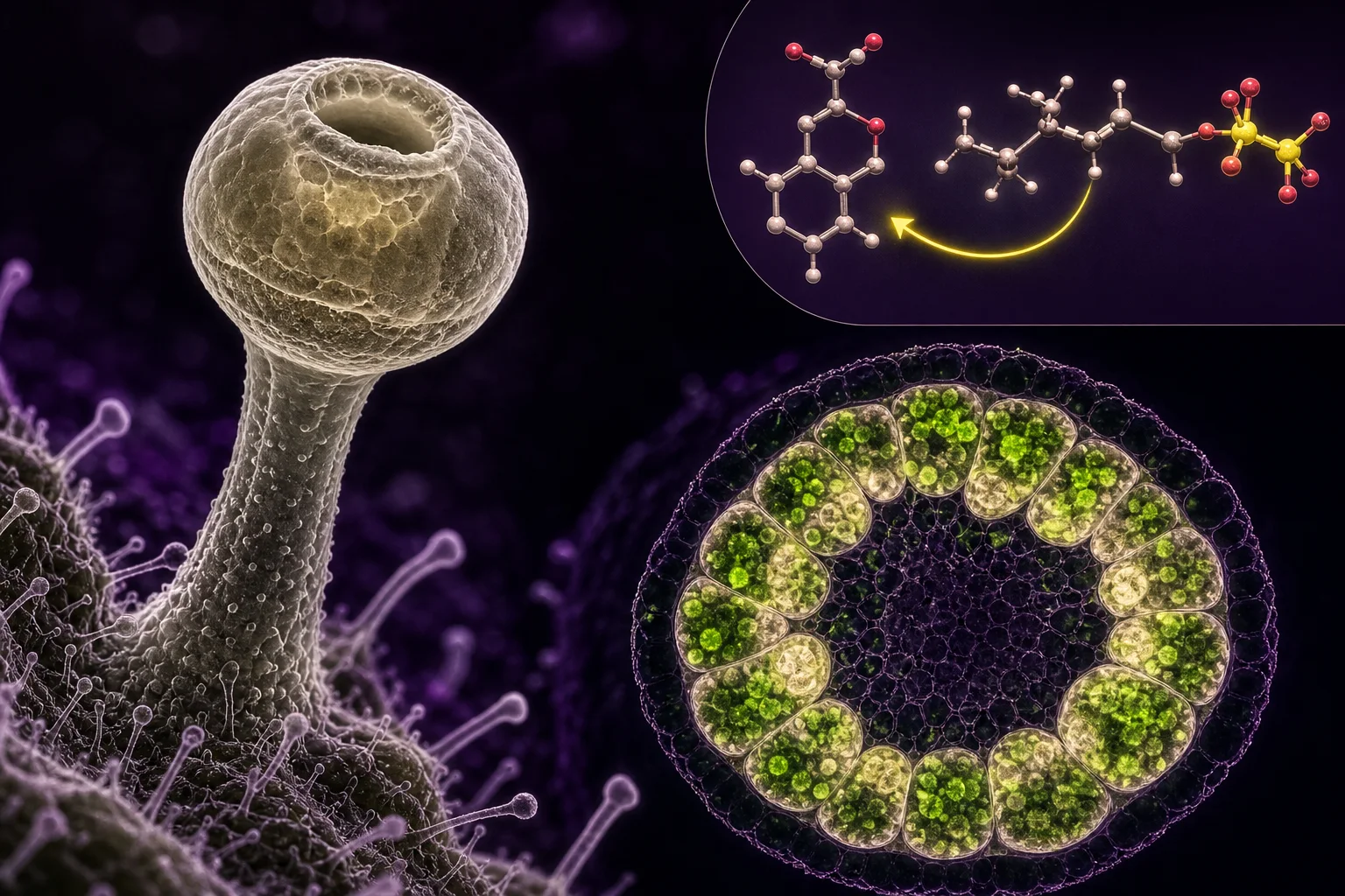
Composite: a single stalked trichome head (microscopy) with plastid-rich disk cells and a schematic molecular inset illustrating OA + GPP → CBGA as a visual key to compartmentalized biosynthesis.
Mainezilla original editorial visual · AI-assisted art directionA short anatomy: the glandular trichome as a production-and-storage unit
Glandular trichomes on female Cannabis flowers are not single simple cells but organized multicellular heads whose apical elements form the metabolite-producing unit. Mature stalked trichomes have a basal stalk and a multicellular head made of a disk of secretory cells and an apical storage (subcuticular) cavity covered by a cuticle. These heads act like tiny microfactories: biosynthetic machinery and subcellular structures are arranged to make, modify, and deposit specialized metabolites into a small extracellular space.
Modern microscopy and transcriptome studies describe the disk as a highly polarized secretory assembly or 'supercell' in which groups of adjacent secretory cells act in a coordinated fashion. That polarization concentrates biosynthetic enzymes and trafficking machinery toward the apical wall that faces the subcuticular cavity, making the head both a site of production and a channel to the extracellular store.
Functionally, it helps to separate synthesis from storage. Lipophilic end products—cannabinoid acids and many terpenes—tend to partition into the cavity, while reactive intermediates and enzymes remain associated with membranes, plastids, or the peripheral cytoplasm around the apical wall. This architecture is central to understanding where a measured compound was likely made and where it is measured.
- Trichome 'heads' = disk of secretory cells + apical subcuticular storage cavity.
- Heads are polarized; many biosynthetic machinery elements concentrate at the apical wall.
- Production (inside cells) and storage (subcuticular cavity) are physically separated.
Plastids: the isoprenoid factory that feeds terpenes (and prenyl groups)
Terpene backbones in plants generally come from two routes for five-carbon building blocks: the cytosolic mevalonate (MVA) route and the plastid-localized methylerythritol phosphate (MEP) route. In Cannabis glandular trichomes the MEP pathway in plastids is the primary supplier of geranyl pyrophosphate (GPP), the ten-carbon donor for monoterpenes and the prenyl donor used in cannabigerolic acid (CBGA) formation.
Electron microscopy and enzyme localization studies find abundant, metabolically active plastids in secretory-stage disk cells. These plastids are non-photosynthetic or lightly pigmented in the mature secretory cells, and they carry the enzymatic steps of the MEP pathway that supply GPP and other isoprenoid intermediates to trichome metabolism.
Because GPP is synthesized inside plastids while other precursors (for example olivetolic acid) are made in the cytosolic/polygonal compartments, the trichome must route those building blocks to the same chemical neighborhood for prenylation. Exactly how plastid-derived GPP is exported or presented to prenyltransferases remains an active research question, but the plastid remains the clear biochemical origin of many terpenoid and prenyl donors in trichomes.
- MEP pathway in plastids supplies GPP (prenyl donor for monoterpenes and CBGA).
- Secretory-stage disk cells contain metabolically active plastids dedicated to specialized metabolism.
- Export/transfer of GPP from plastids to prenylation sites is mechanistically unresolved.
The polyketide pathway and olivetolic acid: making the aromatic core
Cannabinoids begin with an alkylresorcinolic polyketide backbone: olivetolic acid (OA). The core enzymes involved include a type III polyketide synthase (often referred to as tetraketide synthase, TKS) and a partner polyketide cyclase called olivetolic acid cyclase (OAC). Careful trichome transcriptomics and biochemical reconstitution established that OAC is required to convert TKS products into OA.
Crucially, transcriptomic analyses show high expression of the relevant PKS and OAC genes in glandular trichomes, supporting the idea that OA is produced in the secretory cells themselves rather than in distant tissues. Biochemical work that purified and characterized OAC demonstrated a unique aldol condensation mechanism that yields OA with the correct regiochemistry for downstream prenylation.
OA’s synthesis therefore exemplifies the compartmentalized, multi-enzyme choreography inside trichome cells: OA is a polar aromatic intermediate formed in the polyketide pathway, and that intermediate must encounter the plastidial prenyl donor—and the prenyltransferase—to become CBGA, the universal cannabinoid scaffold.
- Type III PKS (TKS) + olivetolic acid cyclase (OAC) synthesize olivetolic acid in trichome cells.
- OAC was identified and structurally characterized; both genes are highly expressed in trichomes.
- OA is the polyketide precursor that must meet plastid-derived GPP to form CBGA.
Prenylation and CBGA: where chemical partners meet
The bond that joins the polyketide OA to the terpenoid prenyl chain is the prenylation step that produces cannabigerolic acid (CBGA). This reaction is catalyzed by an aromatic prenyltransferase (often called GOT/GOT-like or geranylpyrophosphate:olivetolate prenyltransferase), which attaches geranyl diphosphate to the aromatic OA core.
Genetic and subcellular evidence places prenyltransferase activity at membranes and suggests plastid association for at least some prenyltransferases in Cannabis and related plants. A complicating factor is that OA is cytosolic/polyketide-derived and GPP is plastidial, so either the enzyme spans compartments, or metabolite trafficking (for example GPP export or OA import) must bridge those compartments.
Recent syntheses of the cell biology suggest that prenylation likely occurs close to plastids or at membrane contact sites where plastidial GPP is accessible to prenyltransferases. However, the precise membrane topology and exact subcellular address of CBGA formation are not yet settled, and different trichome stages or chemotypes may use subtly different routing.
- CBGA formation = OA + GPP via an aromatic prenyltransferase.
- Prenyltransferases often associate with membranes and can have plastid-targeting signals; trafficking of OA or GPP must bridge compartments.
- Exact subcellular location of prenylation (plastid envelope, ER, or apical face) remains an open question.
THCAS, CBDAS and secretion into the subcuticular cavity
A landmark finding for Cannabis cell biology is that the oxidative cyclases that convert CBGA into the major acidic cannabinoids—THCA synthase (THCAS) and CBDA synthase (CBDAS)—are secreted enzymes. Immunolocalization and biochemical fractionation studies showed THCAS activity and the enzyme itself in the luminal contents of the storage cavity, and fluorescent fusion experiments locate THCAS processing to the trichome head’s secretory apparatus.
This means that some final oxidative steps take place at or beyond the apical cell wall and that the subcuticular cavity is not a passive sink but an active site containing enzymes, substrates, and the products of their reactions. The secretion of THCAS helps explain why acid-form cannabinoids are abundant in the cavity and why analytical sampling of cavity contents returns high levels of THCA or CBDA.
Because THCAS and CBDAS are large, secreted flavoenzymes that can operate on CBGA once released into the cavity environment, the trichome’s architecture (enzymes at the apical face plus a protected extracellular cavity) becomes a crucial kinetic and chemical setting for final product accumulation.
- THCAS and CBDAS are secreted into the subcuticular storage cavity and are active there.
- The storage cavity can contain enzymes, substrates, and final products—effectively an extracellular microreactor.
- Presence of enzymes in the cavity decouples site of final conversion from where intermediates are synthesized.
Terpenes in the headspace and cavity: synthesis and co-accumulation
Terpene synthases expressed in trichome heads create the aromatic bouquet alongside cannabinoid biosynthesis. Many Cannabis terpene synthases are plastidial or plastid-associated, and their products—monoterpenes and sesquiterpenes—are highly lipophilic and readily partition into the same subcuticular cavity that holds cannabinoids.
Advanced microscopic-spectroscopic methods (for example Coherent Anti-Stokes Raman Scattering imaging) have shown spatial co-localization of THCA and selected terpenes inside the secretory cavity at the single-trichome level. That co-localization explains why producers observe consistent chemotype signatures at the trichome level and why one trichome can be chemically complex.
Terpene synthesis, transport, and accumulation interact with cannabinoid deposition: terpene volatility, solubility in the cavity matrix, and possible glycosylation or conjugation pathways influence final profile. Still, plastid origin of many terpenes and the cavity’s role as an amphipathic storage microenvironment are consistently observed across studies.
- Terpene synthases in trichome heads are often plastid-associated and supply monoterpene backbones.
- Microspectroscopic imaging maps terpenes and cannabinoid acids co-localized in the subcuticular cavity.
- Terpene physicochemistry (volatility, partitioning) affects how profiles appear in analyses.
The subcuticular cavity as a chemical microreactor—and what that means for measurements
The subcuticular cavity is small, chemically concentrated, and effectively extracellular. It can contain secreted enzymes (such as THCAS), lipophilic products (THCA, CBDA, terpenes), and reaction microenvironments that differ from intracellular cytosol. For plant physiologists, that means the measured abundance of a compound in a trichome sample reflects both production and partitioning.
For analysts and readers of lab records: techniques that sample whole flower, solvent-rinsed trichomes, or mechanically collected cavity contents will see different relative abundances—acid cannabinoids vs neutral cannabinoids, free terpenes vs glycosylated or bound forms—depending on extraction method and thermal history. For example, GC after heating may decarboxylate acids that HPLC would report intact as THCA/CBDAs.
Mechanistically, a secreted enzyme in the cavity can act on exported intermediates even after cellular secretion—so enzyme localization and simple presence of an intermediate inside the cell are not sufficient to conclude where a compound was formed. Reading claims about 'where' cannabinoids form requires attention to localization data, enzyme secretion evidence, and the physical partitioning documented by microscopic or spectroscopic methods.
- Cavity contents reflect secretion, local enzymatic activity, and partitioning—different from intracellular biosynthesis alone.
- Analytical results depend strongly on sampling and preparation (solvent, heat, mechanical disruption).
- Enzyme location (secreted vs cytosolic) matters for interpreting where conversions happen.
What remains uncertain—and how to read new claims responsibly
Despite rapid progress, important mechanistic gaps remain. The exact membrane topology and subcellular address of the prenyltransferase(s) that make CBGA are not unanimously settled—some data favor membrane-associated plastidal localization, others leave open the possibility of ER or apical-face activity. The transport mechanism(s) that move OA, CBGA, or GPP across membranes or into the cavity (for example dedicated transporters, diffusion via contact sites, or vesicle-mediated routes) remain incompletely described.
Another uncertainty concerns dynamic changes across trichome maturation: the balance between biosynthesis, secretion, and cavity accumulation is time-dependent, and different chemotypes may modulate compartmentation differently. Finally, post-synthetic modifications—glycosylation, acylation, or oxidative rearrangements—are being discovered and may change stability and detectability of certain minor cannabinoids.
When you read a new claim—about a localization, a rate-limiting enzyme, or a novel pathway—check three things: the method (microscopy + immunolocalization or pure proteomics?), the sample preparation (were trichomes intact or disrupted?), and whether the evidence shows activity in situ (enzyme activity in cavity contents or only gene expression?). These contextual details tell you whether a result is cell-biological evidence, an analytical observation, or an inference that needs further functional confirmation.
- Open questions: exact prenyltransferase topology, metabolite transport across membranes, and dynamics during maturation.
- Assess new claims by method (localization vs transcription vs activity), sample prep, and in situ activity evidence.
- Distinguish plant function (mechanism) from analytical detection (what the test measured).
Questions this guide answers
Where in the trichome are cannabinoids first made?
The aromatic polyketide backbone (olivetolic acid) is synthesized in the secretory disk cells via a type III polyketide synthase working with olivetolic acid cyclase; plastids in those same secretory cells supply the geranyl diphosphate prenyl donor. CBGA formation then requires prenylation at membrane-associated sites, and the final oxidative conversions to THCA or CBDA occur after secretion into the subcuticular cavity. Evidence for the polyketide and plastid contributions—and for secreted THCA/CBD enzymes—comes from trichome transcriptomics, enzyme biochemistry, and localization studies. (Sources: 2, 8, 3).
Does THCA form inside the cell or in the cavity?
Key oxidative enzymes (THCAS and CBDAS) have been localized to the apical face of secretory cells and detected as active proteins in the cavity contents; that strongly supports substantial conversion in the cavity or at the apical wall rather than in bulk cytosol. But upstream steps (OA synthesis and prenylation to make CBGA) occur in intracellular compartments, so conversion is distributed across connected microenvironments. (Sources: 3, 4).
If a lab report lists high THCA, does that mean the plant produced it in those trichomes?
High THCA in an analysis means THCA is present in the sampled material, but it does not by itself prove the precise subcellular origin. THCA abundance is influenced by biosynthetic rates, secretion efficiency, partitioning into the cavity, and sample handling before analysis (heat, solvent choice). To infer 'where' it was made, look for corroborating localization or activity assays in the study. (Sources: 6, 3).
Are terpenes made in the same place as cannabinoids?
Many terpene synthases are plastid-associated and produce monoterpenes within or near plastids; because terpenes are lipophilic, they readily partition into the subcuticular cavity alongside cannabinoids. Imaging methods have shown co-localization of certain terpenes and cannabinoid acids in the cavity, but biosynthetic origins (plastid vs polyketide) differ by pathway. (Sources: 5, 6).
Where can I find Maine’s program resources and guidance for editors or growers?
Maine’s Office of Cannabis Policy maintains a resources page with program information, reports, guidance documents and public-health campaigns. It is a useful source for regulatory and editorial context, but not for primary plant-biology mechanisms. (Source: 7).
Educational information only. This guide is not medical or legal advice and does not recommend a product, dose, treatment, or outcome.