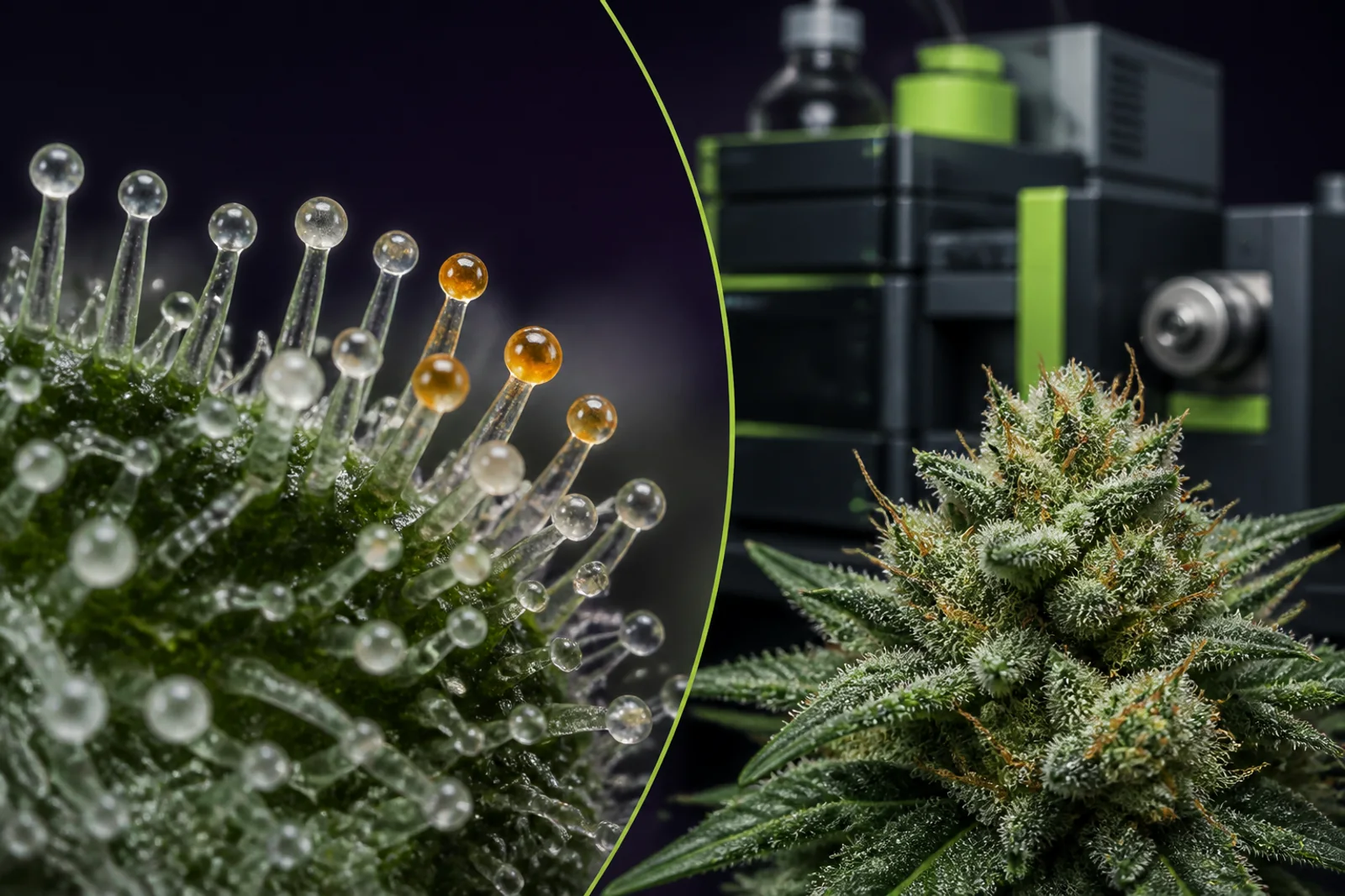
A true macro-style view of glandular trichomes connects visible resin structures to the whole flower and the instruments used to inspect them.
Mainezilla original editorial visual · AI-assisted art directionWhat you are looking at: trichomes in plain language
If you point a macro lens at a ripe cannabis calyx, what looks like frost is a forest of microscopic glands — the glandular trichomes. Those little glassy beads aren’t dust or sugar; they are secretory structures built by the plant to synthesize and store a complex mix of molecules, most notably cannabinoid acids (THCA, CBDA, etc.) and a spectrum of terpenes. Growers call them the plant’s ‘resin factories,’ and that’s literally correct: most of the chemistry we prize in flower happens in those glandular heads. (See evidence: trichome anatomy and cell-type specialization in the literature.)
Not all trichomes are the same. Cannabis has bulbous, sessile capitate, and capitate-stalked glandular trichomes; the capitate-stalked glands on female floral tissues are the heavy hitters for cannabinoids and many terpenes. Bulbous glands tend to be smaller and produce proportionally fewer cannabinoids but can contribute volatiles. Trichome form, density, and developmental trajectory are influenced by genetics and environment; both matter. Growers who recognize which trichomes they’re photographing avoid the ‘what am I even looking at?’ moment when reviewing photos later.
A useful, evidence-forward shorthand: trichomes = production + storage. The secretory disk cells make the molecules and the subcuticular cavity above the disk stores them; over development that cavity can go from clear to milky to amber as contents change. That visible progression is biologically meaningful, but it is not a simple one-to-one proxy for potency or safety without supporting data.
Trichome types and their biology: an essential primer
Capitate-stalked glandular trichomes sit on stalks above the flower surface and have multicellular secretory disks that synthesize cannabinoid acids and many terpenes. As those compounds accumulate the resin builds a subcuticular cavity; microscopy and transcriptomic work show enzyme expression and metabolite accumulation concentrated in these cell types. Researchers isolating trichome heads have repeatedly shown that the biochemical pathways for cannabinoids and terpenes are co-located and often co-regulated, which explains why cultivar chemistry is an integrated trait rather than isolated pockets of chemical behaviour.
Bulbous and sessile trichomes are smaller and have different metabolic outputs; bulbous glands are often more terpene-biased. The distinction matters because a dense-looking surface made up largely of bulbous glands will mean something different chemically than a surface covered in capitate-stalked glands. Developmental stage matters too: trichome density and the composition of stored resin change as flowers mature, and the molecular machinery of the trichome shifts during that maturation.
From a practical perspective, this biology tells us two things: first, trichome appearance is rooted in real, measurable plant processes; second, those processes are complex. You can read clues from morphology and color, but you cannot compute a full chemical profile from a photograph alone. If the goal is a chemical claim—THC percentage, pesticide absence, homogeneity—laboratory testing is the only reliable route.
What a photo reliably shows (and what it doesn’t)
Good photographs can document visual traits: trichome type (stalked vs. sessile), relative density on a particular tissue, the presence of broken or ambering heads, and obvious foreign matter (soil, insects, visible mold). For routine cultivation records, photos act like snapshots in a lab notebook: they’re evidence you can inspect later and compare across rooms, runs, or cultivars. Taken consistently, they reveal trends — a cultivar’s typical trichome density, an environmental problem that reduces gland productivity, or a handling step that bruises resin.
Where photos fail is where they’re most often misused. A crisp macro of milky to amber trichomes does not prove a specific THC percentage, and color alone can’t rule in or out contaminants such as pesticides, heavy metals, mycotoxins, or residual solvents. Lighting, focal depth, magnification, and the surface you chose to photograph (outer bract vs. inner calyx) all change how dense or ambered trichomes appear. Cultivar genetics can make one strain’s amber look another’s ripe: some genetics darken resin pigmentation independent of cannabinoid content.
Put plainly: photos are documentation, not certification. If you want to make a potency claim or to certify an item as microbiologically safe for sale in Maine’s adult-use market, that requires accepted sampling, laboratory analysis, and the state-required Certificate of Analysis. Photographs should be kept beside those lab records — not used to replace them.
How ‘frost’ and ‘ambering’ develop — mechanics and ambiguity
The long-used harvest rules — e.g., wait until a percent of trichomes are milky/amber — reflect real, observable patterns, but they are an oversimplification. Biologically, the shift from translucent to milky to amber corresponds to changes in the composition and oxidation state of the resin stored in the subcuticular cavity, and to structural changes in the trichome head. Transcriptomic and metabolomic studies show that enzymes and metabolites shift during this phase, but the timing and chemistry vary by genetics and environment.
Environmental factors (light spectrum and intensity, temperature swings, humidity, nutrient status) and crop stressors (heat, drought, disease) alter trichome development and resin chemistry. Two cultivars photographed side-by-side under identical handling may show very different trichome color progressions. That’s why some researchers and experienced growers caution against applying a single universal color threshold for harvest across all cultivars — the plant’s genetic program and growth history must be part of the decision.
In short: ambering is a helpful signal in a grower’s toolbox, but it is not a universal certificate of extractability, safety, or therapeutic effect. Treat color as one data point among many — a visual cue that prompts sampling and testing, not a final verdict.
Photographing for records: an evidence-first checklist
If you want photos that will actually help you in hindsight, make them consistent and informative. Minimum data to include with each image: date and time, cultivar or selection name, room or grow location, plant or canopy identifier, plant stage (pre-flower, mid-flower, flush), magnification/optics used, and photographer initials. Store the image next to the matching lab batch number and Certificate of Analysis (COA). A well-labeled photo is comparably useful to a sentence in a logbook; a mystery photo is usually useless.
Photographic best practices: use diffuse, neutral lighting to avoid color cast; include a scale or reference (ruler or coin) for relative size; avoid excessive digital zoom which can blur surface texture; document several tissues (outer bract, inner calyx, sugar leaf) because trichome maturity can vary across them. If you are comparing across runs, keep camera, lighting, and sample prep constant. Minor handling will pop trichome heads; if you squeeze a calyx to get a close shot, note that handling so future reviewers know what they’re seeing.
Labeling and file management matter. Keep a folder structure keyed to batch numbers and testing dates. When paired with lab results and harvest logs, a photo becomes a durable piece of the cultivation record: it helps answer later questions like whether an unexpected lab fail was an isolated handling issue or a recurring cultivation problem.
- Include date, cultivar, room, plant stage, and batch/COA ID with each image.
- Use diffuse lighting, a scale reference, and consistent magnification.
- Photograph multiple tissues; note any handling that could damage trichomes.
Integrating visual cues with Maine’s testing framework
If you cultivate or prepare cannabis that will enter Maine’s licensed adult-use market, understand that the state’s Office of Cannabis Policy (OCP) requires mandatory testing for a set list of analytes. Those analyte categories include THC potency and cannabinoid profiles, water activity, metals, residual solvents and toxins, mycotoxins and harmful microbes, filth/foreign material, and — since phased implementation — pesticides. A visual cue (e.g., ambering) can flag a sampling decision, but passing Maine’s mandatory analyte thresholds requires certified lab testing and a COA issued per OCP rules.
Maine’s testing program also collects and publishes aggregate data on failures and analytes, and the OCP performs audit testing of medical program samples; those audit results have shown a non-trivial prevalence of contaminants in some samples. This is not intended to scare the experienced grower; it is a practical reminder that contaminants are real and not visible under a macro lens. When a photo appears pristine but a lab result fails for pesticides or yeast/mold, it becomes clear why paired documentation (photo + properly taken sample + COA) matters.
Practically: use your photos to trigger sampling (for example, unusual ambering, dust, or visible foreign matter), then follow OCP’s sampling and chain-of-custody guidance and submit timely samples to a certified lab so the final record — your COA — tells the legal and safety story. Photos and COAs together are strong evidence; photos alone are not.
- Maine requires mandatory testing and COAs for adult-use sales; photos do not replace testing.
- OCP publishes testing data and has found contaminants during audit testing.
- Pair visual inspection with proper sampling and certified lab analysis.
Safety and occupational context: contamination, consumer risk, and worker health
There are three safety angles to keep in mind. First, contaminants such as pesticides, heavy metals, and microbial toxins are largely invisible to the camera. Regulators and public health agencies (FDA, CDC) have repeatedly cautioned that products sold outside regulated channels may carry undisclosed contaminants or inaccurate labels. In Maine, OCP’s data and reports document instances where samples would have failed adult-use thresholds if those thresholds had been applied to medical samples, illustrating why testing exists.
Second, different product types carry distinct risks. For example, inhaled products (smoking, vaping, dabbing) have occupational and consumer respiratory considerations — CDC and occupational safety reviews call attention to workplace exposures (dust, fungal spores, volatile organic compounds) and the need for ventilation and exposure controls. Edible products have separate handling and homogeneity concerns; as a legal matter Maine has modified some testing requirements through recent guidance and legislation, underscoring that regulations can change and you should check current OCP guidance before making decisions.
Third, keep records for the sake of traceability and safety. If a consumer or medical patient reports an adverse event, a tidy cultivation folder that includes dated photos, batch IDs, COAs, pesticide application records, and handling logs makes root-cause analysis possible. Treat photographic documentation as part of that traceability chain: it doesn’t prevent every problem, but it helps investigators understand what the plant looked like and when.
Questions this guide answers
Can I tell THC percentage from trichome color alone?
No. Trichome color (clear, milky, amber) is a biological indicator of resin maturation and can inform harvesting decisions, but it doesn’t provide a numeric THC percentage. Accurate potency claims require validated laboratory testing and a Certificate of Analysis.
Do amber trichomes mean the plant is ‘more sedating’?
Not reliably. Traditional lore links ambering with a different balance of cannabinoids and degradation products, but that relationship varies by cultivar and environment. There is no photographic test for subjective effects; evidence-based claims require chemotype profiling and, for clinical claims, clinical research.
If I see no visible mold in photos, can I skip microbial testing?
No. Many microbial risks, including certain yeasts, bacteria, and mycotoxins, are not visible in a macro photograph. Maine’s mandatory testing categories include microbes and water activity for reasons of consumer safety.
How should I use photos in my cultivation records?
Use photos to document date, cultivar, plant or batch ID, room, stage, and sampling events. Store images with your harvest log and COA so they are part of a complete, auditable batch record rather than informal evidence.
Where can I find Maine’s current testing requirements?
Consult the Office of Cannabis Policy (OCP) resources and rule pages for the most recent mandatory testing lists, guidance on sampling, and public testing data. Regulations and guidance have evolved; check OCP’s site for updates.
Educational information only. This guide is not medical or legal advice and does not recommend a product, dose, treatment, or outcome.