
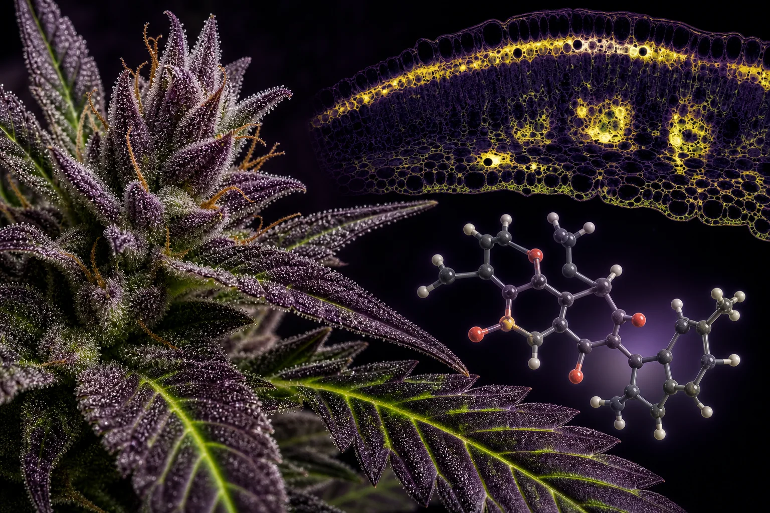
Macro and molecular composite: botanical structure (bract and leaf) overlaid with tissue-level chemical imaging and a flavonoid molecular model to illustrate where flavonoids localize in cannabis tissues.
Mainezilla original editorial visual · AI-assisted art directionWhy tissue localization of flavonoids matters
If you care about plant function or about interpreting a lab sheet, localization is the difference between 'this plant can make a compound' and 'this compound is abundant where we sampled.' Flavonoids in plants serve roles from UV protection and pigmentation to interaction with microbes; their tissue distribution therefore reflects ecological and developmental needs, not marketing categories. ([pmc.ncbi.nlm.nih.gov](https://pmc.ncbi.nlm.nih.gov/articles/PMC10714118/))
For cannabis specifically, a clear separation emerges in the literature between where cannabinoids accumulate (mostly in glandular trichomes on floral bracts) and where flavonoids are biosynthesized and stored, which tends to include leaf tissues and internal floral tissues rather than trichome heads as a primary biosynthetic site. This distinction matters because simple solvent extracts from whole flowers will conflate tissue-level differences into a single number. ([pubmed.ncbi.nlm.nih.gov](https://pubmed.ncbi.nlm.nih.gov/34616415/))
Finally, federal- and state-level testing programs and guidance (including Maine’s Office of Cannabis Policy resources) focus on testing standards, sampling, and lab procedures rather than changing plant chemistry; use those program resources to understand what is being sampled and reported in your jurisdiction. Do not assume program-level data mean the plant molecule is regulated differently. ([maine.gov](https://www.maine.gov/dafs/ocp/home))
Leaves: the flavonoid-rich workshop
Across multiple studies, leaves emerge as a consistent reservoir of flavonoids in Cannabis sativa. Quantitative profiling studies that sampled multiple organs found higher total flavonoid content per dry mass in leaves than in many other tissues, and targeted analyses often detect flavonols (e.g., kaempferol, quercetin derivatives) and glycosides that are classic leaf flavonoids in other plants. These profiles fit a functional story: leaves need UV protection and antioxidant capacity during photosynthesis, so flavonol biosynthesis is a plausible and repeated observation. ([pubmed.ncbi.nlm.nih.gov](https://pubmed.ncbi.nlm.nih.gov/32094454/))
High-resolution mass-spectrometry imaging on leaf surfaces and cross-sections has refined that picture: DESI and MALDI imaging show many flavonoid signals localized within leaf lamina and veins rather than being produced primarily in glandular trichome heads, which have the strongest cannabinoid signals. Imaging thus separates the storage/accumulation compartments of flavonoids from the cannabinoid-rich trichome secretions. Results can vary by cultivar and leaf age, however—so one cultivar’s leaf map is not another’s. ([pubmed.ncbi.nlm.nih.gov](https://pubmed.ncbi.nlm.nih.gov/36654257/))
Practically, that means leaf-based sampling will often show a different flavonoid fingerprint than flower-only samples. If a lab report lists flavonoid totals from ‘whole-plant’ or from leaves specifically, expect higher relative representation of flavonols and glycosides compared with profiles from trichome-rich flower surface extracts. ([pubmed.ncbi.nlm.nih.gov](https://pubmed.ncbi.nlm.nih.gov/32094454/))
Inflorescences and trichomes: a mixed picture
Flowers (inflorescences) are the obvious focus for growers and labs because they host the majority of glandular trichomes, the micro-factories that make and secrete cannabinoids and many terpenoids. Flavonoids are detected in flowers, but their distribution within floral tissues is more heterogeneous: some flavonoids are present within bracts and internal tissues rather than concentrated exclusively in trichome exudate. ([pubmed.ncbi.nlm.nih.gov](https://pubmed.ncbi.nlm.nih.gov/34616415/))
Studies that combine developmental trichome morphology with chemical profiling show that while cannabinoids increase as trichomes mature, flavonoid profiles in the flower can reflect the underlying floral tissue metabolism and developmental timing more than trichome density alone. In short, a flower with dense glands will be cannabinoid-rich, but its flavonoid fingerprint still depends on floral tissue synthesis and timing. ([pmc.ncbi.nlm.nih.gov](https://pmc.ncbi.nlm.nih.gov/articles/PMC10071647/))
Because many laboratories grind whole flowers to prepare extracts, reported floral flavonoid numbers often represent a composite of trichome resin plus internal tissue flavonoids. Interpreting a single floral flavonoid concentration therefore requires asking: Did the sample include bracts, sugar leaves, calyx tissue, or only isolated glandular secretions? Without that context, comparisons across reports are fragile. ([pubmed.ncbi.nlm.nih.gov](https://pubmed.ncbi.nlm.nih.gov/32094454/))
Stems, bark, and roots: understudied but chemically interesting
Comparative tissue surveys show that stems and stem bark generally contain lower total flavonoid concentrations than leaves and flowers, but they are not chemically empty: distinct phenolics, sterols, and specific flavonoids have been quantified in stem tissues and in roots. Roots and subterranean tissues often carry a different signature—more nonpolar phenolics and metabolites associated with belowground functions. ([pubmed.ncbi.nlm.nih.gov](https://pubmed.ncbi.nlm.nih.gov/32094454/))
Cannflavins (A–C and related prenylated flavonoids), first described from flowers, have been detected in multiple organs in different studies, albeit often at lower levels outside leaves and flowers. This suggests that while a flavonoid class may be named from the tissue where it was first isolated, that class can have a broader occurrence across the plant. Concentrations and presence/absence still vary by cultivar and developmental stage. ([pmc.ncbi.nlm.nih.gov](https://pmc.ncbi.nlm.nih.gov/articles/PMC10714118/))
The practical implication for researchers and growers is twofold: (1) expect lower but sometimes unique flavonoid fingerprints in stems and roots, and (2) sampling method matters—peeling bark versus sampling whole stems, or washing and separating root hairs, will influence what the lab detects. Many published datasets do not provide that micro-sampling detail, so interpret with caution. ([pubmed.ncbi.nlm.nih.gov](https://pubmed.ncbi.nlm.nih.gov/32094454/))
Pollen, seeds, and reproductive tissues: data are sparse
Relatively few studies quantify flavonoids in cannabis pollen and seeds compared with leaves and flowers. Available evidence indicates detectable flavonoids in reproductive tissues, but often at low concentrations and with a composition distinct from leaves. Because pollen and seeds serve reproductive and defensive functions, flavonoids there may be tailored to UV protection, pollen–pistil interactions, or antimicrobial defense. ([pmc.ncbi.nlm.nih.gov](https://pmc.ncbi.nlm.nih.gov/articles/PMC10714118/))
When researchers want to study pollen or seed chemistry, sample purity becomes critical. Contamination from adjacent floral tissues or trichome-bearing bracts can inflate flavonoid results if the reproductive material is not carefully isolated. Few published reports include the degree of dissection or cleaning used before analysis, so findings should be read with the same caution applied to stem and root reports. ([pubmed.ncbi.nlm.nih.gov](https://pubmed.ncbi.nlm.nih.gov/32094454/))
Because pollen and seed flavonoids are understudied, they represent a small but important gap where targeted, tissue-purified studies could reveal distinct molecules with ecological or biological relevance—especially across male vs. female plants in this dioecious species. ([pmc.ncbi.nlm.nih.gov](https://pmc.ncbi.nlm.nih.gov/articles/PMC12861918/))
Developmental stage, plant sex, and genetics shape flavonoid maps
Flavonoid biosynthesis is developmentally regulated. Time-course and gene-expression studies in cannabis show that key biosynthetic genes vary in expression across maturation of leaves and inflorescences, producing different flavonoid fingerprints as organs age and as flowering progresses. Chromatin and transcriptional work also show epigenetic control that can change flavonoid production independently of cannabinoid pathways. ([pmc.ncbi.nlm.nih.gov](https://pmc.ncbi.nlm.nih.gov/articles/PMC12861918/))
Sex differences in cannabis (male vs. female plants) matter because floral morphology and trichome distribution differ; gene-expression studies report intersexual differences in both secondary metabolite pathways and trichome-related genes. Put simply: the same cultivar grown as a male or a female—or sampled at week 3 versus week 6 of flowering—can show different flavonoid patterns. ([pubmed.ncbi.nlm.nih.gov](https://pubmed.ncbi.nlm.nih.gov/41994336/))
Genetic background (chemovar) remains a major source of variation. Multiple profiling studies across chemovars and growth conditions demonstrate that cultivar-specific enzymatic complements and regulation produce divergent flavonoid suites. That is why a single lab value from one cultivar shouldn’t be generalized across all cannabis. ([pubmed.ncbi.nlm.nih.gov](https://pubmed.ncbi.nlm.nih.gov/32094454/))
How methods change the map: imaging, extraction, and analytical caveats
Analytical method shapes the story you get. Imaging mass spectrometry (DESI, MALDI) maps compounds in situ and can distinguish laminar vs. surface localization, whereas bulk extraction followed by LC-MS or spectrophotometry reports averaged concentrations across the mixture. Imaging studies show flavonoid signals in leaf tissue while trichome heads show dominant cannabinoid signals—an important methodological distinction. ([pubmed.ncbi.nlm.nih.gov](https://pubmed.ncbi.nlm.nih.gov/36654257/))
Extraction solvent, grinding, and whether sugar leaves or bracts were co-sampled directly alter reported flavonoid levels. Glycosides and aglycones differ in solubility; some laboratories hydrolyze conjugates before analysis while others measure intact glycosides, producing different reported profiles. Always check a report’s sample description and the analytical method (e.g., targeted LC-MS vs. total flavonoid colorimetric assays). ([pubmed.ncbi.nlm.nih.gov](https://pubmed.ncbi.nlm.nih.gov/32094454/))
Finally, consider limits of detection, standardization, and inter-lab differences. Because many flavonoid standards are expensive or unavailable, labs sometimes quantify relative to surrogate standards or report semi-quantitative results. That means a minor flavonoid reported at low micrograms per gram in one lab might be below detection in another—compare methods, not just numbers. ([pubmed.ncbi.nlm.nih.gov](https://pubmed.ncbi.nlm.nih.gov/32094454/))
Reading claims, records, and what still needs work
When you see a single flavonoid concentration on a certificate of analysis (COA), ask: which tissue was sampled, what was the cultivar, what extraction and quantitation method was used, and when in the life cycle was the sample taken? Many differences in published flavonoid prevalence come down to those four variables. Program resources like Maine’s Office of Cannabis Policy explain testing frameworks and can help you understand how sampling protocols may influence reported results. ([maine.gov](https://www.maine.gov/dafs/ocp/home))
Uncertainties remain. We still lack broad, multicultivar, multi-site time-course datasets that match cell-level localization (imaging) with bulk analytics and transcriptomics across organs and sexes. Key open questions include the exact cellular compartments for most flavonoid biosynthetic enzymes in cannabis and the ecological drivers of tissue-specific flavonoid expression across diverse cultivars. ([pmc.ncbi.nlm.nih.gov](https://pmc.ncbi.nlm.nih.gov/articles/PMC12861918/))
For practitioners and readers of reports: treat flavonoid numbers as cultivar- and context-specific. Use them to compare like-with-like (same tissue, same developmental stage, same method), and flag comparisons that do not meet those criteria as provisional. That approach keeps botanical respect for variability at the center of interpretation. ([pubmed.ncbi.nlm.nih.gov](https://pubmed.ncbi.nlm.nih.gov/32094454/))
Questions this guide answers
Are flavonoids produced in trichomes the same as those in leaves?
Not necessarily. Trichome secretions are dominated by cannabinoids and terpenoids; imaging and tissue-specific studies show many flavonoids are synthesized in leaf lamina and internal floral tissues rather than concentrated in trichome heads. However, some flavonoids are present in flowers at detectable levels and may be partially associated with surface tissues after grinding or extraction. ([pubmed.ncbi.nlm.nih.gov](https://pubmed.ncbi.nlm.nih.gov/36654257/))
Do roots and stems contain useful amounts of flavonoids?
Roots and stems typically have lower total flavonoid concentrations than leaves and flowers but can contain distinct phenolic and flavonoid signatures. Their profiles are understudied and can include compounds with different polarities and functions compared with aerial tissues. Sampling technique strongly affects measured levels. ([pubmed.ncbi.nlm.nih.gov](https://pubmed.ncbi.nlm.nih.gov/32094454/))
Can I compare flavonoid numbers from two different labs?
Only if the labs used comparable sampling (same tissue and developmental stage), extraction, and analytical methods. Differences in standards, hydrolysis/non-hydrolysis of glycosides, and limits of detection make many inter-lab comparisons unreliable unless method details are aligned. ([pubmed.ncbi.nlm.nih.gov](https://pubmed.ncbi.nlm.nih.gov/32094454/))
Does Maine regulate flavonoids differently than cannabinoids?
Maine’s Office of Cannabis Policy provides regulatory resources and testing frameworks focused on safety, labeling, and testing standards. It does not regulate individual plant molecules in a way that changes their biology; regulatory attention is on testing practices, sampling, and public health considerations rather than altering where flavonoids naturally occur in the plant. Consult OCP resources for program context. ([maine.gov](https://www.maine.gov/dafs/ocp/home))
What are the biggest gaps for researchers?
Large, systematic, multi-chemovar studies that combine tissue-purified imaging, transcriptomics/epigenomics, and standardized bulk analytic chemistry across timepoints are lacking. Such datasets would help resolve where flavonoid biosynthesis occurs at the cellular level and how genetics and environment interact to shape tissue-specific flavonoid distributions. ([pmc.ncbi.nlm.nih.gov](https://pmc.ncbi.nlm.nih.gov/articles/PMC12861918/))
Educational information only. This guide is not medical or legal advice and does not recommend a product, dose, treatment, or outcome.